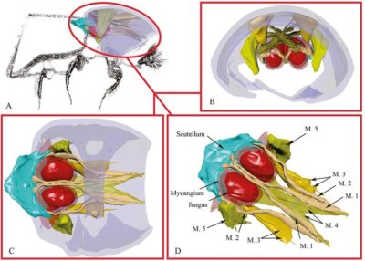
In our constant quest to understand the anatomy and behavior of ambrosia beetles, our lab decided to experiment with 3D imaging techniques that could reveal the details of mycangia.
Mycangia are anatomical structures that most species of ambrosia beetles have in one form or another which allow them to carry samples of the fungus they depend on entirely for food from one tree host to another. They are typically complex hollows in the body of the beetle. Understanding the structures of the mycangia can help us to zero in on what species of beetle and fungi might eventually become invasive pests that threaten forests.
Entomology Today thought that our imaging experiments might be helpful to other entomologists who are considering new ways of examining tiny structures. So at their request, Dr. Jiri Hulcr (our PI) and Jackson Landers (our Strategic Science Communicator) authored an article describing our experiences. Read all about it and watch the video online.
